


Inside Aesthetics
The Crows Nest
USA

Clinical Defects and the Technical Approach
Woodrow Wilson and Dario Bertossi

INTRODUCTION TO THE TECHNICAL DEVICES FOR HIGHLIGHTING AND REPORTING CLINICAL DEFECTS: PHOTOGRAPHY AND OPTICAL IMAGING
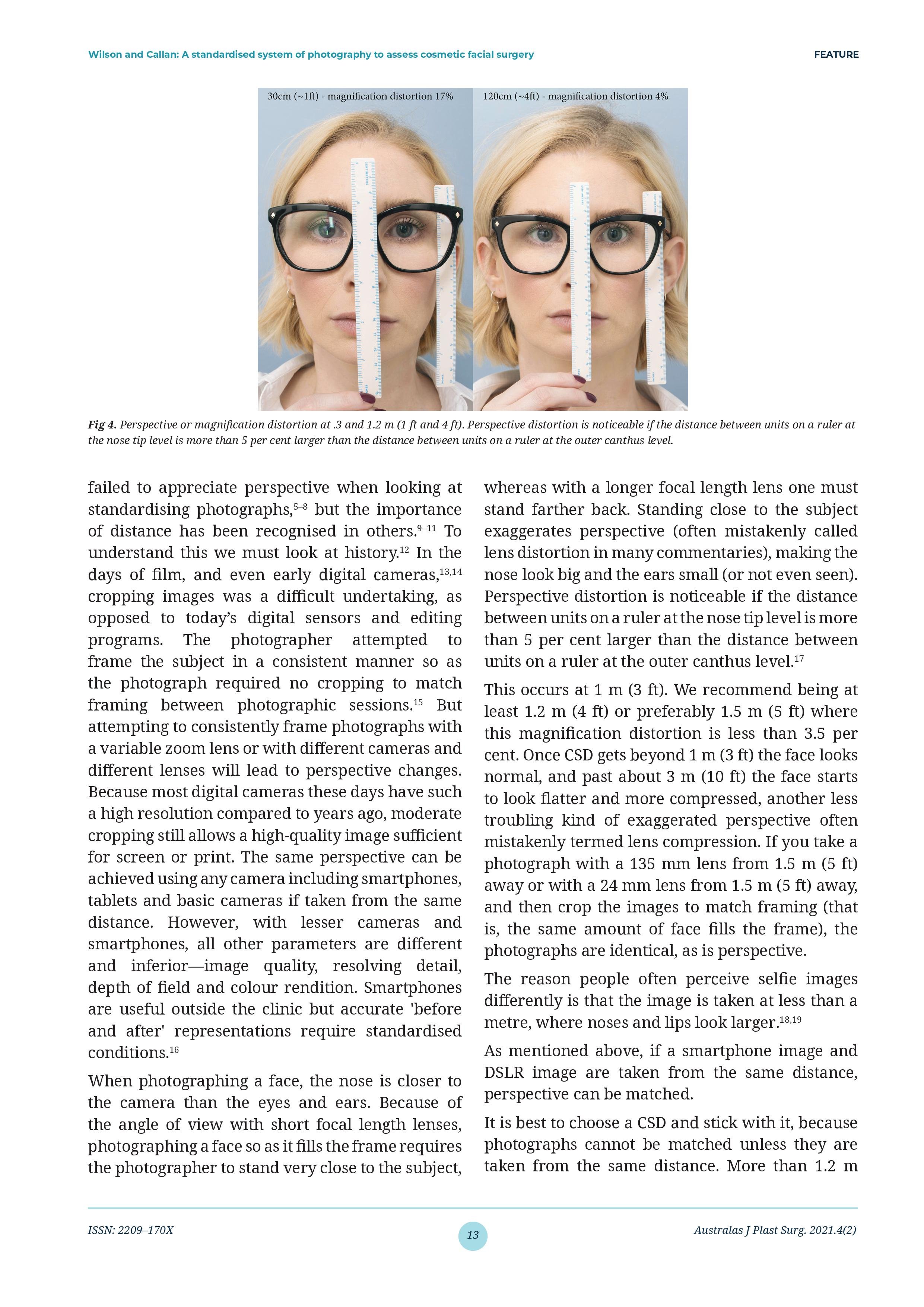
Surgeons and physicians engaged in plastic surgery and aesthetic medicine should rely on a scientific objective and accurate imaging data of patients undergoing the treatment, just starting from optical photography [1]. There are three technological pathways for capturing clinical photography—traditional two-dimensional (2D) using a digital single lens reflex (DSLR), three-dimensional (3D) surface imaging or augmented reality. 3D imaging has come to have a big impact on aesthetic surgery worldwide [1,2]. The first attempt to use the 3D surface imaging technique in a clinic dates to 1944 by Thalmaan, who used stereo photogrammetry to examine an adult with facial asymmetry and a baby with Pierre Robin syndrome.
Since then, photography for aesthetic medicine evolved, and 3D photography has become greatly used, allowing for a more dynamic facial evaluation, although it is associated with an increased cost [3,4]. Accurate 2D imaging as a lower-cost entry point has been pursued [13]. Doctors approaching either plastic surgery or non-surgical rhinoplasty or profileplasty should be ensured of the soundness and high-quality outcome of patients’ photos, as the advantages of a high sharpness in the visual result of a photo allow the surgeon to screen and detect any small defects on the face skin and anatomy, enabling him or her to plan a correct surgical or non-surgical intervention [5]. The principles outlined in this chapter allow for accurate capture of the nose, but also the entire face, mandible and neckline.
Continue at DOI: 10.1201/9781003304623-12
A standardised system of photography to assess cosmetic facial surgery
Callan PP, Wilson W. A standardised system of photography to assess cosmetic facial surgery. AJOPS. 2021;4(2):8-21. doi:10.34239/ajops.v4n2.334


Injections in Facial Aesthetic Medicine An International Key Expert Guide

Philipp-Dormston_De Boulle_ Swift_Almeida_Seo_Flynn_
Abstract
Clinical photography has never been more important, prevalent or misunderstood. Whilst clinical photography in plastic surgery has a long history of attempted standardisations, aesthetic practitioners performing injectable treatments has grown along with the rise in smartphone technology as the primary photographic capture device, for both clinicians and patients. As smartphone photographs cannot be standardised easily, both clinicians and patients are unwittingly capturing anatomy which is not objectively accurate.
This chapter looks to educate the reader on the technology, novel protocols and justifications for optimal use of digital photography in an aesthetic clinic. We will systematically examine all the variables and share standards for any clinician to use, regardless of technical prowess. A common misconception is that you need to be a photographer to perform excellent clinical photography. In reality you simply need standardised equipment, protocol and motivation.
My aim is to instil an appreciation for the invaluable nature of an objective, scientific and reproducible approach to clinical photography - not only as a vital tool for documentation and education of your patient, but as a valuable business asset in an increasingly competitive environment. We will discuss systematic steps to avoid technological bias introduced through poor photographical methodology, and present robust techniques and setup parameters.
While the quality of smartphone cameras are always improving and do have their place in intraoperative capturing. It is my firm experience that they cannot provide the level of standardisation that is required to perform accurate clinical photography.
Publication due 2025